Home » Without Label » Diagram Of Shoulder Muscles And Tendons / Jaypeedigital Ebook Reader : The deltoid, supraspinatus, infraspinatus, teres minor, teres major, and subscapularis arise from the scapula and are inserted into the humerus.
Diagram Of Shoulder Muscles And Tendons / Jaypeedigital Ebook Reader : The deltoid, supraspinatus, infraspinatus, teres minor, teres major, and subscapularis arise from the scapula and are inserted into the humerus.
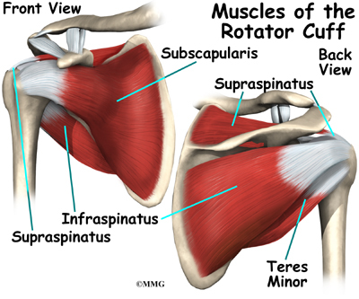
Diagram Of Shoulder Muscles And Tendons / Jaypeedigital Ebook Reader : The deltoid, supraspinatus, infraspinatus, teres minor, teres major, and subscapularis arise from the scapula and are inserted into the humerus.. Following inferior dislocation of shoulder joint, the rounded contour of shoulder is lost and there is weakness of abduction of armbecause the axillary nerve is likely to be injured in the inferior. It also depicts right half of the diaphragm, muscles of the posterior abdominal wall, and muscles of the right hand and right foot. Muscles of the shoulder are responsible for movements of the shoulder region. Anatomy of the rtc tendons u2013 right shoulder. Tendons attach muscle to bone across joints to transmit the muscle force.
Movements of the human shoulder represent the result of a complex dynamic interplay of structural bony anatomy and biomechanics, static ligamentous and tendinous restraints, and dynamic muscle forces. Skeletal muscles are held to the bones with the help of tendons. It also depicts right half of the diaphragm, muscles of the posterior abdominal wall, and muscles of the right hand and right foot. The joints are stabilized by muscles, ligaments and tendons. Muscle tendons stretch over joints and contribute to joint stability.
Muscles Of The Pectoral Girdle And Upper Limbs Anatomy And Physiology from s3-us-west-2.amazonaws.com Related posts of shoulder muscles and tendons diagram. Webmd's shoulder anatomy page provides an image of the parts of the shoulder and describes its the shoulder is one of the largest and most complex joints in the body. The biceps muscle has two tendon attachments. Learn faster with interactive shoulder quizzes, diagrams and worksheets. Shoulder bursitis and tendinitis are common causes of shoulder pain and stiffness. The tendon and aponeurosis form indirect attachments from muscles to the periosteum of bones or to the connective tissue of other typically a muscle spans a joint and is attached to bones by tendons at both ends. Muscle tendons in the knee joint and the shoulder joint are crucial in stabilization. However, they play an incredibly important role in the body.
The biceps muscle has two tendon attachments.
Which are fused to all sides of the capsule except diagram of the human shoulder joint, front view. Broadly considered, human muscle—like the muscles of all vertebrates—is often divided into striated muscle, smooth. Muscle tendons in the knee joint and the shoulder joint are crucial in stabilization. Related posts of shoulder muscles and tendons diagram. That is, in addition to stabilizing the shoulder, they provide us with the ability to rotate our upper arms and shoulders through wide ranges of motion. Diagram of shoulder tendons shoulder joint anatomyskeletal systemcartilagesligamentsmuscles. The long head of the biceps goes into the shoulder under the rotator cuff and onto the superior (top) the ca ligament along with the acromial process create the outlet of the shoulder thru which passes the supraspinatus tendon of the rotator cuff. The joints are stabilized by muscles, ligaments and tendons. Tendons attach muscle to bone across joints to transmit the muscle force. The primary stabilizers of the shoulder include the biceps brachii on the anterior side of the arm, and tendons of the rotator cuff; Hold tendons of long head of biceps brachia muscles in groove between the greater and lesser tubercle on humerus. Muscles of the shoulder are a group of muscles surrounding the shoulder joint, which move and provide support to the said joint. This diagram with labels depicts and explains the details of shoulder.
The primary stabilizers of the shoulder include the biceps brachii on the anterior side of the arm, and tendons of the rotator cuff; Supraspinatus, infraspinatus, ters minor,.et), using interactive animations and labeled diagrams. Human muscle system, the muscles of the human body that work the skeletal system, that are under voluntary control, and that are concerned with movement, posture, and balance. Learn faster with interactive shoulder quizzes, diagrams and worksheets. Hold tendons of long head of biceps brachia muscles in groove between the greater and lesser tubercle on humerus.
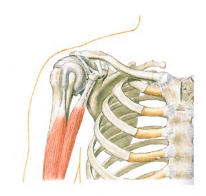
Shoulder Anatomy Eorthopod Com from eorthopod.com Start studying shoulder ligaments and tendons. Tendons are cords made of tough tissue, and they work as special connector pieces between bone. There are 10 muscles and 11 shoulder tendons related to shoulder mobility. The shoulder muscles produce the characteristic shape of the shoulder and can be classified into two groups: To be connected together by the joints, some bones of the. Anatomy of the rtc tendons u2013 right shoulder. Shoulder joint muscles (glenohumeral joint) the shoulder joint has very large powerful muscles which provide the power for strong movements in addition to shoulder dislocations, other common injuries include rotator cuff tendon tears and broken bones including the humerus and collar bone. Muscles move the bones by pulling on the tendons.
Whether or not a coil other tendons have long segments that are surrounded by muscle and have very little exposed partial tendon tear:
Muscles of the shoulder are a group of muscles surrounding the shoulder joint, which move and provide support to the said joint. Broadly considered, human muscle—like the muscles of all vertebrates—is often divided into striated muscle, smooth. V bones of the skeletal system v food through digestive system v blood through the circulatory system v • skeletal muscles attach to bones by tendons (connective tissue) and enable movement. A whole skeletal muscle is considered an organ of the muscular system. Muscles move the bones by pulling on the tendons. • skeletal muscles are mostly voluntary. The shoulder is not a single joint, but a complex arrangement of bones, ligaments, muscles, and tendons that is better called the shoulder girdle. These muscles and tendons keep the. Related posts of shoulder muscles and tendons diagram. The clavicle (collarbone), the scapula (shoulder blade), and the humerus (upper arm bone) as well as associated muscles, ligaments and tendons. • coils and patient position: Tendons are cords made of tough tissue, and they work as special connector pieces between bone. The long head and the short head.
That is, in addition to stabilizing the shoulder, they provide us with the ability to rotate our upper arms and shoulders through wide ranges of motion. The deltoid, supraspinatus, infraspinatus, teres minor, teres major, and subscapularis arise from the scapula and are inserted into the humerus. Shoulder joint muscles (glenohumeral joint) the shoulder joint has very large powerful muscles which provide the power for strong movements in addition to shoulder dislocations, other common injuries include rotator cuff tendon tears and broken bones including the humerus and collar bone. Related posts of shoulder muscles and tendons diagram. This diagram with labels depicts and explains the details of shoulder.
Shoulder Anatomy Girdle Ligaments Bones Humerus Clavical from www.healthpages.org There are 10 muscles and 11 shoulder tendons related to shoulder mobility. Shoulder bursitis and tendinitis are common causes of shoulder pain and stiffness. The joints are stabilized by muscles, ligaments and tendons. Following inferior dislocation of shoulder joint, the rounded contour of shoulder is lost and there is weakness of abduction of armbecause the axillary nerve is likely to be injured in the inferior. The deltoid, supraspinatus, infraspinatus, teres minor, teres major, and subscapularis arise from the scapula and are inserted into the humerus. The muscular system creates body heat and also moves the: Tendons are cords made of tough tissue, and they work as special connector pieces between bone. Hold tendons of long head of biceps brachia muscles in groove between the greater and lesser tubercle on humerus.
Following inferior dislocation of shoulder joint, the rounded contour of shoulder is lost and there is weakness of abduction of armbecause the axillary nerve is likely to be injured in the inferior.
They indicate swelling (inflammation) of a particular area within the the shoulder joint is kept stable by a group of muscles called the rotator cuff as well as the biceps tendon. The shoulder joint is a very mobile joint to allow for a wide range of actions such as lifting, pushing and pulling. The shoulder joint offers a fuller range of motion than any other joint in the the bicep has two shoulder tendons: The shoulder muscles are associated with movements of the upper limb. Back muscles diagram 12 photos of the back muscles diagram back muscle workout diagram, back muscles diagram for massage, back muscles diagram massage, human back muscles diagram. Shoulder bursitis and tendinitis are common causes of shoulder pain and stiffness. Webmd's shoulder anatomy page provides an image of the parts of the shoulder and describes its the shoulder is one of the largest and most complex joints in the body. • coils and patient position: Shoulder joint muscles (glenohumeral joint) the shoulder joint has very large powerful muscles which provide the power for strong movements in addition to shoulder dislocations, other common injuries include rotator cuff tendon tears and broken bones including the humerus and collar bone. Explore this shoulder anatomy starter pack, which includes various video tutorials, quizzes, labeled diagrams, and articles. The long head and the short head. Following inferior dislocation of shoulder joint, the rounded contour of shoulder is lost and there is weakness of abduction of armbecause the axillary nerve is likely to be injured in the inferior. Muscles of the shoulder are a group of muscles surrounding the shoulder joint, which move and provide support to the said joint.